Part 1 – Intro | Part 2 – Coronary Heart Disease | Part 3 – Angina | Part 4 – Heart Attack
What goes wrong – More on the Pathology of Coronary Heart Disease (CHD)
“Diabetes is a major risk factor for heart disease…”
“Heart Disease is twice as common in people with diabetes…”
What goes wrong? Why are people with diabetes more prone to heart disease?
This page addresses heart function in more detail, and looks at some of the underlying causes of coronary heart disease, with special consideration towards the effects of diabetes.
What’s covered on this page
Normal Heart Functioning – How the Heart Should Work
Atherosclerosis – Hardening of the Arteries
Coronary Heart Disease (CHD)
Diagnostic Tests and Procedures
Normal Heart Functioning – How the Heart Should Work
Introduction to How the Heart Works
Firstly, let’s review the basic structure and function of the heart. The heart is about the size of a mans’ clenched fist and is situated in the chest between the lungs. It pumps blood around the body, supplying oxygen and nutrients essential for life. It is largely muscle, which contracts and relaxes (‘beats’) continually, day and night.
Blood is carried away from the heart in arteries. Arterial blood is rich in oxygen. The blood arrives at various organs and tissues around the body, and the cells in these tissues use up the oxygen. Veins carry blood back to the heart. Venous blood is low in oxygen.
The oxygen that we need to survive comes from the air that we breathe. It is taken into the body through the lungs. The heart and lungs work together to take the oxygen to the body’s cells.
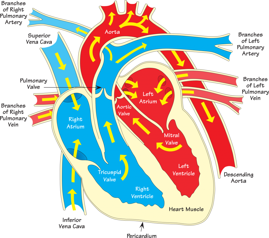
The heart has four ‘chambers’, two on each side. The right side of the heart collects ‘used’ blood from the body, and then pumps it to the lungs. The blood, now rich in oxygen, returns from the lungs to the left side of the heart. The left side of the heart then pumps the oxygen-rich blood to the rest of the body. The left and right pairs of chambers consist of a small atrium (also called “auricle”), which collects blood, and a larger ventricle, which pumps blood out of the heart. The blood flow is controlled by the heart valves, which only allow blood through in one direction.
|
Figure 1 The Heart
 |
The Cardiac Cycle
Blood that is returning from the various body organ systems empties into the right atrium from two large veins. The inferior vena cava delivers blood from parts of the body below the heart. The superior vena cava delivers blood from above the heart.
Inside the top of the right atrium lies a bundle of muscle and nerve fibres – this is the pacemaker, otherwise known as the sinoatrial node, SA node. At regular intervals an electrical signal is fired from here, which causes the muscles of the right atrium to contract, forcing the blood down through a one-way valve, and into the right ventricle. The SA node determines the rate at which the heart beats.
As the chamber fills with blood the electrical signal from the SA node reaches another bundle of muscle and nerve fibres – the atrioventricular node or AV node – which lies at the bottom of the atrium. The AV node picks up the electrical impulse and spreads it across the heart. The ventricle is stimulated to contract and blood is forced out through the pulmonary artery towards the lungs.
The left atrium and left ventricle respond almost simultaneously to the waves of electrical signals. The left atrium collects oxygen-rich blood from the lungs through the pulmonary veins. The left ventricle then pumps this blood out through the aorta, to deliver oxygen to the rest of the body.
The entire process of emptying and re-filling the heart lasts less than one second.
| The period when the chambers are in relaxation phase, allowing blood to flow in, is called DIASTOLE.
The period of contraction of the chamber walls, forcing blood through the one-way valves, is termed SYSTOLE. The three phases that together make a heartbeat are therefore:
|
The characteristic sounds made by the blood rushing through the various valves in the heart can be heard with the use of the stethoscope.
Atherosclerosis or “Hardening of the Arteries”
Normal healthy arteries have strong but flexible walls, and a smooth inner lining (the endothelium). However, the inner lining can become laden with fatty materials such as cholesterol, other lipids, calcium and other materials. Soft fatty deposits ultimately become thicker, hardened and make areas of the artery stiff and narrowed. Such lesions, called plaque, clog the artery in much the same way as rust, grease and scale can block the drain in the kitchen sink.
|
Terminology Check Atherosclerosis usually refers to damage to the innermost lining of the arterial wall. Arteriosclerosis is a term that is sometimes used in a broader sense to include calcification of the middle layer of the artery wall. |
Atherosclerosis occurs over a long period of time. It is a painless process in itself, and we are largely unaware that our arteries are becoming clogged up – until other symptoms of arterial disease present, that is.
People with diabetes are more likely to develop atherosclerosis. Other risk factors for atherosclerosis include high levels of blood cholesterol, smoking, high blood pressure, a sedentary lifestyle, and being overweight.
Consequences of Atherosclerosis
Atherosclerosis can potentially affect any of the body’s arteries – for example those supplying the brain, neck, arms, legs, kidneys, lungs, and of course, the heart.
| Atherosclerosis causes plaque to form on the lining of arteries, narrowing these blood vessels and reducing blood flow to an area of the body.
Patches of atheroma or “plaques” gradually build up, and as they do so they thicken and weaken the walls of the affected artery. If the plaque tears or ruptures a clot (“thrombus“) may form. |
Thrombosis
Normal healthy arteries are smooth and elastic. However, when ‘hardening of the arteries’ occurs, the inside surface becomes rough, uneven and breaks may occur, encouraging platelets and other blood cells to stick together in theory to form a clot that seals the break. The protrusion of the clot into the blood vessel may be enough to significantly reduce blood flow. This is frequently the cause of pain in “unstable angina” if this happens in a coronary artery.
Fatty deposits can become surrounded by scar tissue and a rigid fibrous cap may form over the lesion. Damage to the fibrous cap can lead to the formation of a large clot, which may completely block the artery. When a clot blocks the vessel, the blood flow is stopped, and the area of tissue that the artery is supplying will be starved of oxygen and nutrients; the tissue beyond the clot is seriously damaged and the consequences can be far-reaching. A stroke may result if this occurs in a cerebral artery in the neck or brain, or a heart attack may result if it occurs in a coronary artery supplying the heart itself. If it occurs in the trunk of the body it may lead to an aneurysm. If a limb is affected, the process may lead to gangrene.
Atherosclerosis in peripheral arteries – in the legs, for example – causes peripheral arterial disease (PAD). This is an important feature of diabetes complications and is considered in detail in the section, “legs and feet“
Atherosclerosis, or ‘hardening of the arteries’, in the coronary arteries causes coronary heart disease (CHD), sometimes referred to as coronary artery disease (CAD) (see below).
Coronary Heart Disease (CHD)
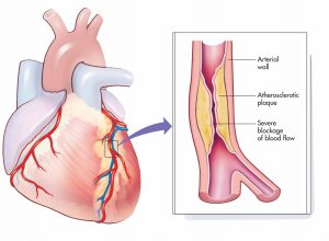
The coronary arteries are the blood vessels that supply the heart muscle itself with ‘fresh’ oxygenated blood. In order for the heart to work properly on a continuing basis the heart muscle requires a good blood supply. Any damage to the arteries that supply the heart – i.e. the coronary arteries – can limit the blood supply to the heart muscle, and if the damage eventually causes the artery to become blocked then it may turn out to be life-threatening.
|
Figure 2 The heart, showing origin of coronary artery disease
|
A deficiency of blood to the heart muscle is called ischaemia. It may be “silent” (no symptoms, not noticed), or it may cause significant chest pain, which we refer to as ‘angina’ (see Part 3). Silent ischaemia is more common in people with long-standing diabetes – damage to the nervous system can impair the perception of pain, and in some cases this may render people with diabetes at higher risk of suffering a ‘surprise’ heart attack.
The human body has designed its own backup in reserve for blocked coronary arteries; the ‘collateral circulation‘ is a system of small blood vessels that offer an alternative route between two segments of the same artery, or sometimes between two arteries. The blockage can therefore sometimes be by-passed, if the appropriate alternative route for blood flow is available via the collateral circulation. However, this alternative circulation is not well developed in everyone, and compensation may not be sufficient to sustain normal heart function.
| There is evidence to suggest that the growth of collateral vessels may be stimulated through exercise. |
The net effect of coronary heart disease is generally to reduce the amount of blood that the heart muscle receives. This in turn may lead to angina or heart attack.
Diagnostic Tests and Procedures
| Angiography is the most widely used technique used to visualise blood vessels in the body.
Ultrasound can be used to assess some arteries – those in the neck and thighs, for example. CT or CAT scanning (Computed tomography) is sometimes used to detect calcium deposits in blood vessels. PET scanning (positron-emission tomography) may be used to determine if a tissue or organ has an adequate blood supply. An ECG (electrocardiogram or EKG) records the electrical activity of the heart. The heart produces tiny electrical impulses which spread through the heart muscle to make the heart contract. These impulses can be detected by the ECG machine. The ECG is commonly used to detect abnormal heart rhythms and to investigate the cause of chest pain. Stress-tests or exercise ECG tests may be used for:
|

